
 Electron Micrographs


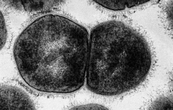
(70,000X)
High magnification electron micrograph
of an ultra-thin section of a group A streptococcus sibling pair. At this magnification,
especially in the cell on the left, the cell wall and cell surface fibrils,
consisting primarily of M protein, are well
defined. Interdigitaion of these fibrils between neighboring cells of different
chains is also in plain view.
Strain: C126/21/1;
M-type 43.