 Electron Micrographs

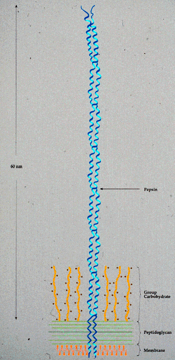
Model of Streptococcal M Protein
The coiled-coil dimeric nature of M protein and its relationship to the bacterial cell surface is shown. The N-terminal region of the M protein, distal to the cell surface, varies among different M types, thereby providing the molecular basis of Dr. Lancefield's method of serotyping group A streptococci. In contrast, the C-terminal region of M protein, commencing at the pepsin susceptible site, is more conserved. The physical relationship between the cell membrane, cell wall, the group A specific carbohydrate and M protein are also indicated.
For a recent review of the biology of the streptococcal M protein, see :
Fischetti, V. A. 1991. Streptococcal M protein. Scientific American. 264:58-65.


