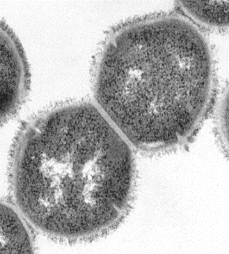
 Electron Micrographs
(16,000X)
Electron micrograph of an ultra-thin section of two group A streptococci from a chain of cells. The septum between the two cells is clearly indicated by the light colored diagonal line in the center of the image. The bacterial chromosome is also clearly seen as the light staining material in the cell interior. Fibrils on the cell surface contain the type-specific M protein characteristic of S. pyogenes.


