
 Electron Micrographs


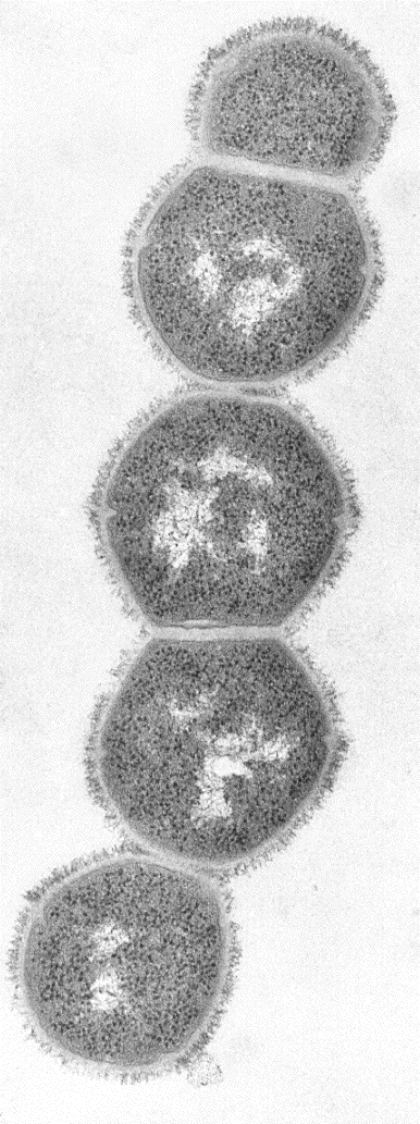
(20,000X)
Electron micrograph of an ultra-thin section of a chain of group A streptococci. The cell surface fibrils, consisting primarily of M protein, are clearly evident. The bacterial cell wall, to which the fibrils are attached, is also clearly seen as the light staining region between the fibrils and the dark staining cell interior. Incipient cell division is also indicated by the nascent septum formation (seen as an indentation of the cell wall) near the cell equator. The streptococcal cell diameter is equal to approximately one micron.